![Medical News Asia | Powered by ['mediPr]](https://medi-paper.com/wp-content/uploads/2020/02/MediPR-MediPaper-Medical-Communications-Ltd-Healthcare-Writers-Medical-Writers-Medical-Writer-Agency-Hong-Kong-Medical-Writing-Services-Solutions-Quote-醫學寫作-香港-醫學作家香港-medical-PR-public-relations-1030x374.png)
HER2 Explorer
Navigating HER2 Testing: Unveiling the Strengths and Limitations to Optimize Biomarker Assessment
Last updated: 31 July 2025 | In: HER2 Explorer, Breast Cancer
Introduction
Due to the availability of human epidermal growth factor receptor 2 (HER2)-directed therapies, testing for HER2 alterations, including overexpression, gene amplification or mutations is recommended in treatment guidelines for various tumor types, including breast cancer, gastric cancer, biliary tract cancer, cervical cancer, colorectal cancer, non-small cell lung cancer (NSCLC), and others.1-101. National Comprehensive Cancer Network. NCCN Clinical Practice Guidelines in Oncology – Biliary Tract Cancers Version 1.2025.
2. National Comprehensive Cancer Network. NCCN Clinical Practice Guidelines in Oncology – Bladder Cancer Version 1.2025.
3. National Comprehensive Cancer Network. NCCN Clinical Practice Guidelines in Oncology – Ovarian Cancer Including Fallopian Tube Cancer and Primary Peritoneal Cancer Version 1.2025.
4. National Comprehensive Cancer Network. NCCN Clinical Practice Guidelines in Oncology – Cervical Cancer Version 4.2025.
5. National Comprehensive Cancer Network. NCCN Clinical Practice Guidelines in Oncology – Colon Cancer Version 3.2025.
6. National Comprehensive Cancer Network. NCCN Clinical Practice Guidelines in Oncology – Rectal Cancer Version 2.2025.
7. National Comprehensive Cancer Network. NCCN Clinical Practice Guidelines in Oncology – Uterine Neoplasms Version 3.2025.
8. National Comprehensive Cancer Network. NCCN Clinical Practice Guidelines in Oncology – Non-Small Cell Lung Cancer Version 3.2025.
9. National Comprehensive Cancer Network. NCCN Clinical Practice Guidelines in Oncology – Gastric Cancer Version 2.2025.
10. National Comprehensive Cancer Network. NCCN Clinical Practice Guidelines in Oncology – Breast Cancer Version 4.2025.
To ensure patients eligible to receive HER2-directed therapies are identified, accurate assessment of HER2 status is essential. The guideline-recommended testing modality varies depending on the type of specific HER2 alteration present, such as overexpression, gene amplification or mutations, for which a HER2-directed therapy is available in specific tumor types. For example, HER2 immunohistochemistry (IHC) testing, which detects HER2 surface protein expression, is recommended by National Comprehensive Cancer Network (NCCN) guidelines for a variety of solid tumors.4,7,9,104. National Comprehensive Cancer Network. NCCN Clinical Practice Guidelines in Oncology – Cervical Cancer Version 4.2025.
7. National Comprehensive Cancer Network. NCCN Clinical Practice Guidelines in Oncology – Uterine Neoplasms Version 3.2025.
9. National Comprehensive Cancer Network. NCCN Clinical Practice Guidelines in Oncology – Gastric Cancer Version 2.2025.
10. National Comprehensive Cancer Network. NCCN Clinical Practice Guidelines in Oncology – Breast Cancer Version 4.2025. For a detailed review of HER2 IHC scoring, please refer to Article 3 of this series. For NSCLC, guidelines recommend HER2 IHC testing for overexpression, and next-generation sequencing (NGS) to evaluate HER2 mutations.88. National Comprehensive Cancer Network. NCCN Clinical Practice Guidelines in Oncology – Non-Small Cell Lung Cancer Version 3.2025. Where NGS is not available, Sanger sequencing or polymerase chain reaction (PCR) assays are acceptable alternatives.88. National Comprehensive Cancer Network. NCCN Clinical Practice Guidelines in Oncology – Non-Small Cell Lung Cancer Version 3.2025. Furthermore, NGS is also capable of detecting HER2 gene amplification, and it can detect multiple variants across the genome, including novel or rare genetic variants, and evaluate gene copy number.11-1311. Illumina. Advantages of NGS vs Sanger sequencing. Available online. https://sapac.illumina.com/science/technology/next-generation-sequencing/beginners/advantages/ngs-vs-sanger.html. Last accessed: 2 December 2024.
12. Illumina. Differences between NGS and qPCR. Available online.https://sapac.illumina.com/science/technology/next-generation-sequencing/beginners/advantages/ngs-vs-qpcr.html. Last accessed: 2 December 2024.
13. Niu D, et al. Pathol Oncol Res. 2020;26:2577–85.
KEY FEATURES of HER2 testing modalities
Currently, multiple companion diagnostic assays for HER2 evaluation have been cleared for use by the US Food and Drug Administration (FDA), including options for IHC, fluorescent in situ hybridization (FISH) and NGS panel (Table 1).1414. US Food and Drug Administration. List of Cleared or Approved Companion Diagnostic Devices (In Vitro and Imaging Tools). Available online.https://www.fda.gov/medical-devices/in-vitro-diagnostics/list-cleared-or-approved-companion-diagnostic-devices-in-vitro-and-imaging-tools. Last accessed: 2 December 2024.
Each testing modality targets specific biomolecular features, and while there may be overlap in certain capabilities, such as detecting gene amplifications, their methodologies and outputs offer distinct clinical insights (Table 2).11,12,15-1811. Illumina. Advantages of NGS vs Sanger sequencing. Available online. https://sapac.illumina.com/science/technology/next-generation-sequencing/beginners/advantages/ngs-vs-sanger.html. Last accessed: 2 December 2024.
12. Illumina. Differences between NGS and qPCR. Available online.https://sapac.illumina.com/science/technology/next-generation-sequencing/beginners/advantages/ngs-vs-qpcr.html. Last accessed: 2 December 2024.
15. Loong HH, et al. JTO Clin. Res Rep. 2022;3(3):100290.
16. Kamps R, et al. Int J Mol Sci. 2017;18:(2):308.
17. Zalis M, et al. Front Genet. 2024;15:1420190.
18. Furrer D, et al. Am J Clin Pathol. 2015;144(5):686–703.
- IHC (Immunohistochemistry): Used to evaluate HER2 protein expression on the cell surface, IHC remains a widely adopted initial diagnostic tool due to its cost-efficiency and standardized protocols. However, variability in antibody performance and subjective interpretation can influence consistency.
- FISH (Fluorescence In Situ Hybridization): Primarily employed to assess HER2 gene amplification by quantifying copy number directly, FISH offers clarity when IHC results are equivocal. Its targeted nature lends precision, but it is typically slower and more resource-intensive.
- NGS (Next-Generation Sequencing) Panels: NGS enables simultaneous profiling of various genetic alterations—including point mutations, fusions, and gene amplifications such as HER2. It delivers a broader genomic snapshot compared to FISH, making it well-suited for more comprehensive molecular characterization. However, NGS does not provide insight into protein levels and requires higher-quality tissue input.
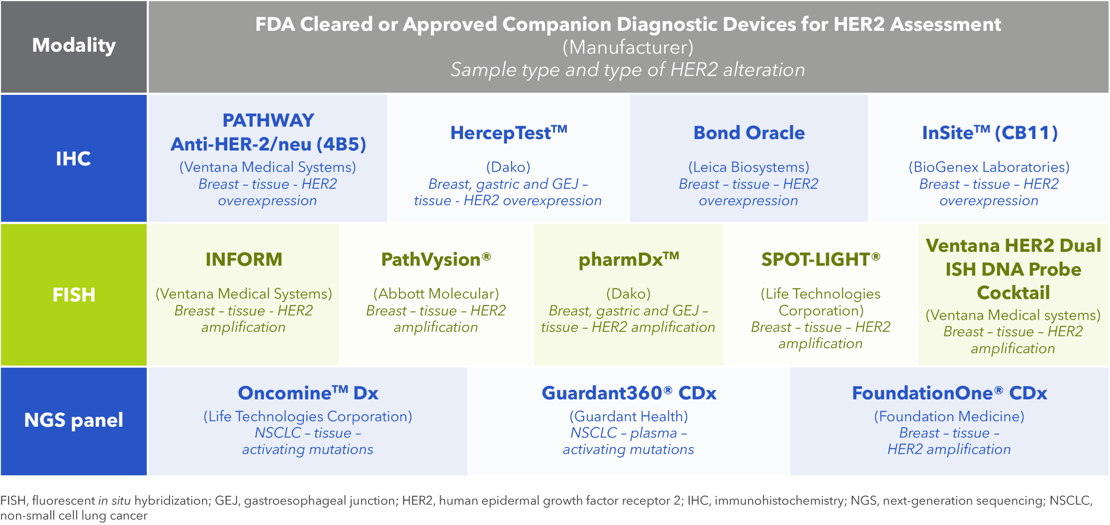
Table 1. FDA-cleared diagnostic devices for the evaluation of HER2 status in solid tumors1414. US Food and Drug Administration. List of Cleared or Approved Companion Diagnostic Devices (In Vitro and Imaging Tools). Available online.https://www.fda.gov/medical-devices/in-vitro-diagnostics/list-cleared-or-approved-companion-diagnostic-devices-in-vitro-and-imaging-tools. Last accessed: 2 December 2024.
This table is not intended to directly compare the advantages and disadvantages of IHC, FISH, and NGS panels, as each method assesses distinct types of HER2 alterations. Rather than being viewed in isolation, these tests should be considered complementary, providing a more comprehensive evaluation when used together.
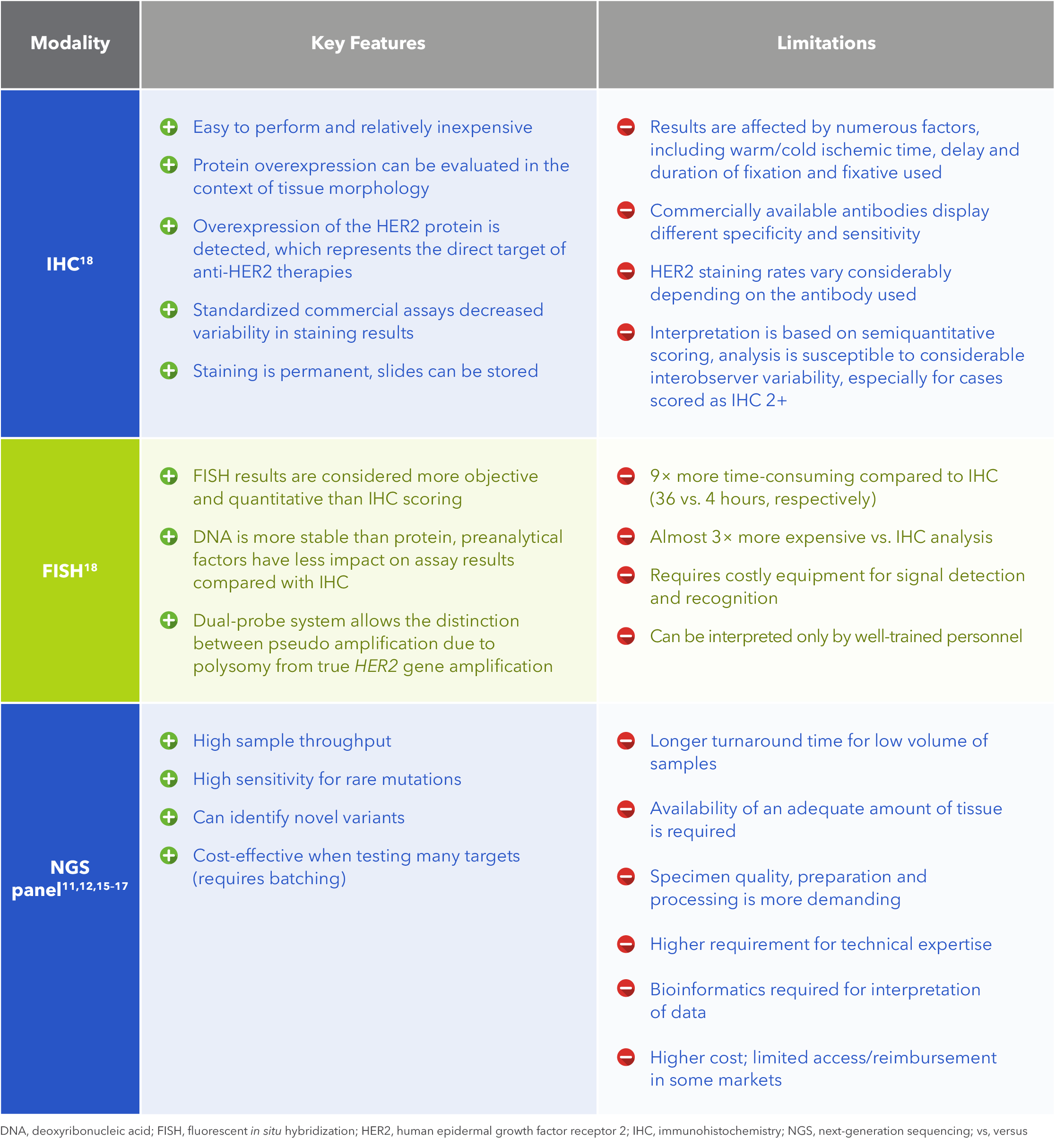
Table 2. Features and limitations of IHC, FISH and NGS panel for assessment of HER2 status11,12,15-1811. Illumina. Advantages of NGS vs Sanger sequencing. Available online. https://sapac.illumina.com/science/technology/next-generation-sequencing/beginners/advantages/ngs-vs-sanger.html. Last accessed: 2 December 2024.
12. Illumina. Differences between NGS and qPCR. Available online.https://sapac.illumina.com/science/technology/next-generation-sequencing/beginners/advantages/ngs-vs-qpcr.html. Last accessed: 2 December 2024.
15. Loong HH, et al. JTO Clin. Res Rep. 2022;3(3):100290.
16. Kamps R, et al. Int J Mol Sci. 2017;18:(2):308.
17. Zalis M, et al. Front Genet. 2024;15:1420190.
18. Furrer D, et al. Am J Clin Pathol. 2015;144(5):686–703.
Standardization strategies to minimize HER2 assessment variability
The accuracy of HER2 assays may be adversely impacted by various factors. Accurate and reproducible testing is essential to ensure that false-negative and false-positive results are minimized.1919. Ivanova M, et al. Virchows Arch. 2024;484(1):3–14. Clinicians should be aware of the guideline specifications for time to formalin fixation and the recommended duration, as delays to fixation or under/over fixation may affect deoxyribonucleic acid (DNA) quality for NGS testing and impact IHC/FISH results.20-2220. Wolff AC, et al. J Clin Oncol. 2018;36(20):2105–22.
21. Yamashita-Kashima Y, et al. Gastric Cancer. 2014;17(4):638–47.
22. Arreaza G, et al. Int J Mol Sci. 2016;17(19):1579.
Best practice in tissue preparation to improve HER2 IHC/FISH testing accuracy
Careful management of pre-analytical issues is crucial for both biopsy and surgical specimens to minimize the occurrence of false-negative and false-positive results.1919. Ivanova M, et al. Virchows Arch. 2024;484(1):3–14. With regards to biopsy or surgical excision, the American Society of Clinical Oncology/College of American Pathologists (ASCO/CAP) guidelines for HER2 IHC/FISH testing in breast cancer recommend cold ischemia time be minimized to ≤1 hour.19,20,2319. Ivanova M, et al. Virchows Arch. 2024;484(1):3–14.
20.Wolff AC, et al. J Clin Oncol. 2018;36(20):2105–22.
23. Wolff AC, et al. J Clin Oncol. 2023;41(22):3867–72. Subsequent testing should be performed on formalin-fixed paraffin-embedded (FFPE) tissue samples derived from primary, recurrent or metastatic tissues.19,2319. Ivanova M, et al. Virchows Arch. 2024;484(1):3–14.
23. Wolff AC, et al. J Clin Oncol. 2023;41(22):3867–72. Furthermore, tissue samples for HER2 testing should be fixed in 10% neutral buffered formalin for 6–72 hours, and cytology specimens must be fixed in formalin.2323. Wolff AC, et al. J Clin Oncol. 2023;41(22):3867–72. After paraffin embedding, samples should be sliced at 5–10 mm intervals after appropriate gross inspection, margins designation and being placed in sufficient volume of neutral buffered formalin.2323. Wolff AC, et al. J Clin Oncol. 2023;41(22):3867–72. Any exceptions to this process must be included in the HER2 test report.2323. Wolff AC, et al. J Clin Oncol. 2023;41(22):3867–72. Sections should ideally not be used for HER2 testing if they are >6 weeks old, however, this may vary with primary fixation or storage conditions.2020. Wolff AC, et al. J Clin Oncol. 2018;36(20):2105–22. To prevent discrepancies in results interpretation, laboratories should use validated HER2 assays and participate in regular laboratory inspection proficiency training.2020. Wolff AC, et al. J Clin Oncol. 2018;36(20):2105–22. Workflow and operating procedures were standardized as an example to optimize HER2 testing in breast cancer, particularly for the assessment of HER2-low status (Figure 1).1919. Ivanova M, et al. Virchows Arch. 2024;484(1):3–14.
Best practice in tissue preparation to improve HER2 NGS testing accuracy
Expert opinion on biomarker testing for small specimens in NSCLC recommends the use of core needle biopsy, bronchoscopic forceps biopsy, and fine-needle aspiration, with or without endobronchial ultrasound (EBUS), for obtaining tissue samples.2424. Penault-Llorca F, et al. Virchows Arch. 2022;481(3):335–50. However, if tissue samples are insufficient or impractical to obtain, cytological samples and liquid biopsy may serve as appropriate alternatives.2424. Penault-Llorca F, et al. Virchows Arch. 2022;481(3):335–50. To ensure reliable genetic alteration testing, at least ~10% of the sample should consist of neoplastic cells.2424. Penault-Llorca F, et al. Virchows Arch. 2022;481(3):335–50. Macro-microdissection may be necessary to enrich samples for NGS analysis.2424. Penault-Llorca F, et al. Virchows Arch. 2022;481(3):335–50.
The NCCN guidelines recommend rapid on-site evaluation (ROSE) to confirm the adequacy of transbronchial needle aspirates (TBNAs) and EBUS specimens for testing.2424. Penault-Llorca F, et al. Virchows Arch. 2022;481(3):335–50. Once collected, tissue processing should be optimized to maximize DNA quality.2424. Penault-Llorca F, et al. Virchows Arch. 2022;481(3):335–50. In particular, unbuffered formalin or fixatives containing acids or heavy metals should be avoided.2424. Penault-Llorca F, et al. Virchows Arch. 2022;481(3):335–50. Instead, tissue should be fixed in 10% neutral buffered formalin for 6–48 hours.2424. Penault-Llorca F, et al. Virchows Arch. 2022;481(3):335–50.
In cases of bone biopsies, decalcification should only be performed as a last resort using ethylenediaminetetraacetic acid (EDTA).2424. Penault-Llorca F, et al. Virchows Arch. 2022;481(3):335–50. Additionally, to minimize tissue wastage, fragments should be separated into individual blocks following a ‘one sample per block’ approach.2424. Penault-Llorca F, et al. Virchows Arch. 2022;481(3):335–50. This strategy reduces material loss that would otherwise occur due to the need to create a single plane encompassing multiple tissue fragments.2424. Penault-Llorca F, et al. Virchows Arch. 2022;481(3):335–50.
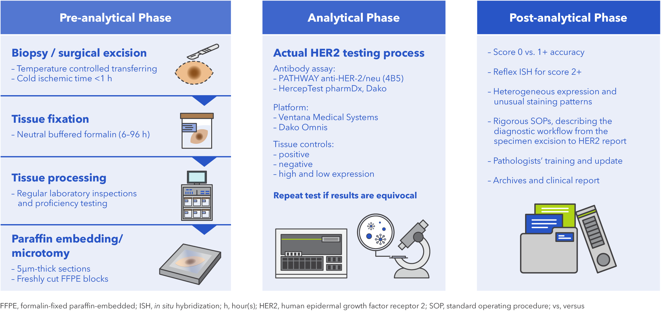
Figure 1. Suggested standard operating procedure for optimization of HER2-low status assessment1919. Ivanova M, et al. Virchows Arch. 2024;484(1):3–14.
Addressing inter-pathologist and inter-antibody discordances in HER2 IHC assessment
Although the standardization of tissue handling and laboratory techniques can reduce the variability of HER2 testing results, discordance among pathologists when assessing IHC samples is another potential source of variability.2525. Robbins CJ, et al. Mod Pathol. 2023;36(1):100032. A study of 170 breast cancer biopsies by 18 specialized pathologists from 15 institutions identified discordance within the intermediate categories (Figure 2).2525. Robbins CJ, et al. Mod Pathol. 2023;36(1):100032. The study demonstrated that using a simplified three-category scoring system (IHC 0, intermediate, or 3+) improved overall agreement from 28.8% to 46.5% compared to the traditional four-category system. Importantly, while interpathologist discordance was observed, concordance was substantially higher when the task was limited to distinguishing HER2 IHC 3+ from non–3+ cases. In this context, the overall percent agreement (OPA) plateaued at 87.1% with six raters, indicating that pathologists are more consistent when identifying cases with strong HER2 overexpression.2525. Robbins CJ, et al. Mod Pathol. 2023;36(1):100032.
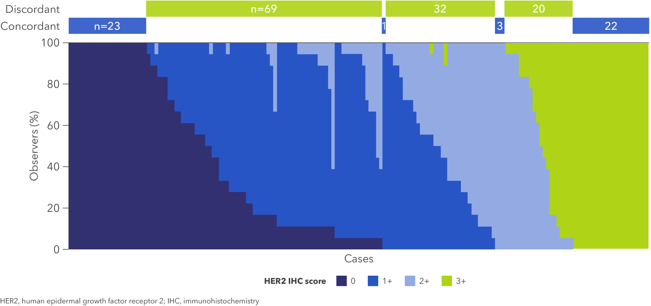
Figure 2. HER2 IHC score for breast cancer specimens (n=170) assigned by 18 pathologists2525. Robbins CJ, et al. Mod Pathol. 2023;36(1):100032.
Differences between antibody clones used in IHC assays are another potential source of discordance.2626. Karakas C, et al. Am J Clin Pathol. 2023;159(5):484–91. An analysis of 114 breast cancer specimens scored by six pathologists according to ASCO/CAP breast cancer guidelines compared results between the HercepTest (Agilent Dako, polyclonal anti-HER2 antibody) and PATHWAY (Ventana, 4B5 anti-HER2 antibody) IHC assays.2626. Karakas C, et al. Am J Clin Pathol. 2023;159(5):484–91. The overall interantibody agreement rate was 57.8%, with higher agreement rates seen among IHC 2–3+ specimens (91.9% HercepTest, 86.3% PATHWAY) than among IHC 0–1+ (78.1% HercepTest, 72.2% PATHWAY) (Figure 3).2626. Karakas C, et al. Am J Clin Pathol. 2023;159(5):484–91. Although discordance between antibody clones exists, particularly in HER2-low or negative cases, strong concordance is observed among pathologists when identifying higher levels of HER2 expression.
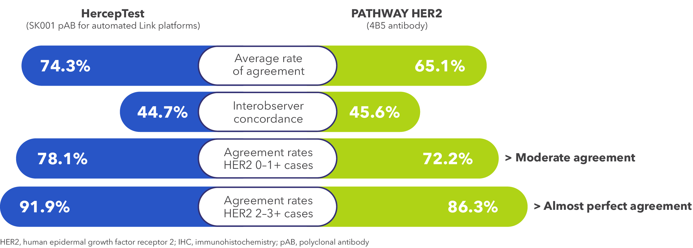
Figure 3. Summary of agreement rates of HER2 IHC scores between HercepTest (polyclonal anti-HER2 antibody) and PATHWAY (4B5 anti-HER2 antibody) in 114 breast cancer specimens evaluated by six pathologists2626. Karakas C, et al. Am J Clin Pathol. 2023;159(5):484–91.
Conclusions
Accurate assessment of HER2 status is essential to identify patients with HER2 overexpression, gene amplification or mutations, who are eligible for HER2-directed therapies. Various modalities of HER2 testing are recommended by guidelines.1-101. National Comprehensive Cancer Network. NCCN Clinical Practice Guidelines in Oncology – Biliary Tract Cancers Version 1.2025.
2. National Comprehensive Cancer Network. NCCN Clinical Practice Guidelines in Oncology – Bladder Cancer Version 1.2025.
3. National Comprehensive Cancer Network. NCCN Clinical Practice Guidelines in Oncology – Ovarian Cancer Including Fallopian Tube Cancer and Primary Peritoneal Cancer Version 1.2025.
4. National Comprehensive Cancer Network. NCCN Clinical Practice Guidelines in Oncology – Cervical Cancer Version 4.2025.
5. National Comprehensive Cancer Network. NCCN Clinical Practice Guidelines in Oncology – Colon Cancer Version 2.2025.
6. National Comprehensive Cancer Network. NCCN Clinical Practice Guidelines in Oncology – Rectal Cancer Version 2.2025.
7. National Comprehensive Cancer Network. NCCN Clinical Practice Guidelines in Oncology – Uterine Neoplasms Version 3.2025.
8. National Comprehensive Cancer Network. NCCN Clinical Practice Guidelines in Oncology – Non-Small Cell Lung Cancer Version 3.2025.
9. National Comprehensive Cancer Network. NCCN Clinical Practice Guidelines in Oncology – Gastric Cancer Version 2.2025.
10. National Comprehensive Cancer Network. NCCN Clinical Practice Guidelines in Oncology – Breast Cancer Version 3.2035.
Together with optimized handling of samples and implementation of standardized operating procedures, pathology labs can minimize rates of false-positive and false-negative assays.1919. Ivanova M, et al. Virchows Arch. 2024;484(1):3–14. Discordance among pathologists or between different anti-HER2 antibodies used in breast cancer IHC tests have been reported, however, agreement rates remain high with IHC 3+ cases.25,2625. Robbins CJ, et al. Mod Pathol. 2023;36(1):100032.
26. Karakas C, et al. Am J Clin Pathol. 2023;159(5):484–91. Pathologists should stay current with evolving biomarker evaluation criteria and reporting standards, as the concept of the HER2 spectrum expands with the inclusion of IHC 0+ in the latest CAP report template update.19,27-2919. Ivanova M, et al. Virchows Arch. 2024;484(1):3–14.
27. Tozbikian G, et al. Arch Pathol Lab Med. 2024;148(2):242–55.
28. Venetis K, et al. Front Mol Biosci. 2022;9:834651.
29. 29. College of American Pathologists. Reporting Template for Reporting Results of Biomarker Testing of Specimens from Patients with Carcinoma of the Breast. Available online. https://documents.cap.org/documents/New-Cancer-Protocols-March-2025/Breast.Bmk_1.6.0.0.REL.CAPCP.pdf. Last accessed: 23 May 2025. Further development of more accurate and reproducible testing methods is warranted optimize HER2 testing and improve the accuracy of patient identification.19,2619. Furrer D, et al. Am J Clin Pathol. 2015;144(5):686–703.
26. Karakas C, et al. Am J Clin Pathol. 2023;159(5):484–91.
References
- National Comprehensive Cancer Network. NCCN Clinical Practice Guidelines in Oncology – Biliary Tract Cancers Version 1.2025.
- National Comprehensive Cancer Network. NCCN Clinical Practice Guidelines in Oncology – Bladder Cancer Version 1.2025.
- National Comprehensive Cancer Network. NCCN Clinical Practice Guidelines in Oncology – Ovarian Cancer Including Fallopian Tube Cancer and Primary Peritoneal Cancer Version 1.2025.
- National Comprehensive Cancer Network. NCCN Clinical Practice Guidelines in Oncology – Cervical Cancer Version 4.2025.
- National Comprehensive Cancer Network. NCCN Clinical Practice Guidelines in Oncology – Colon Cancer Version 3.2025.
- National Comprehensive Cancer Network. NCCN Clinical Practice Guidelines in Oncology – Rectal Cancer Version 2.2025.
- National Comprehensive Cancer Network. NCCN Clinical Practice Guidelines in Oncology – Uterine Neoplasms Version 3.2025.
- National Comprehensive Cancer Network. NCCN Clinical Practice Guidelines in Oncology – Non-Small Cell Lung Cancer Version 3.2025.
- National Comprehensive Cancer Network. NCCN Clinical Practice Guidelines in Oncology – Gastric Cancer Version 2.2025.
- National Comprehensive Cancer Network. NCCN Clinical Practice Guidelines in Oncology – Breast Cancer Version 4.2025.
- Illumina. Advantages of NGS vs Sanger sequencing. Available online. https://sapac.illumina.com/science/technology/next-generation-sequencing/beginners/advantages/ngs-vs-sanger.html. Last accessed: 2 December 2024.
- Illumina. Differences between NGS and qPCR. Available online.https://sapac.illumina.com/science/technology/next-generation-sequencing/beginners/advantages/ngs-vs-qpcr.html. Last accessed: 2 December 2024.
- Niu D, et al. Pathol Oncol Res. 2020;26:2577–85.
- US Food and Drug Administration. List of Cleared or Approved Companion Diagnostic Devices (In Vitro and Imaging Tools). Available online.https://www.fda.gov/medical-devices/in-vitro-diagnostics/list-cleared-or-approved-companion-diagnostic-devices-in-vitro-and-imaging-tools. Last accessed: 2 December 2024.
- Loong HH, et al. JTO Clin. Res Rep. 2022;3(3):100290.
- Kamps R, et al. Int J Mol Sci. 2017;18:(2):308.
- Zalis M, et al. Front Genet. 2024;15:1420190.
- Furrer D, et al. Am J Clin Pathol. 2015;144(5):686–703.
- Ivanova M, et al. Virchows Arch. 2024;484(1):3–14.
- Wolff AC, et al. J Clin Oncol. 2018;36(20):2105–22.
- Yamashita-Kashima Y, et al. Gastric Cancer. 2014;17(4):638–47.
- Arreaza G, et al. Int J Mol Sci. 2016;17(19):1579.
- Wolff AC, et al. J Clin Oncol. 2023;41(22):3867–72.
- Penault-Llorca F, et al. Virchows Arch. 2022;481(3):335–50.
- Robbins CJ, et al. Mod Pathol. 2023;36(1):100032.
- Karakas C, et al. Am J Clin Pathol. 2023;159(5):484–91.
- Tozbikian G, et al. Arch Pathol Lab Med. 2024;148(2):242–55.
- Venetis K, et al. Front Mol Biosci. 2022;9:834651.
- College of American Pathologists. Reporting Template for Reporting Results of Biomarker Testing of Specimens from Patients with Carcinoma of the Breast. Available online. https://documents.cap.org/documents/New-Cancer-Protocols-March-2025/Breast.Bmk_1.6.0.0.REL.CAPCP.pdf. Last accessed: 23 May 2025.
The article is sponsored by Daiichi Sankyo and AstraZeneca.
MED-HK-TRAS-00099 HK-11852 07/2025
Disclaimer
This article is not medical advice. Patients should seek personal assessment by a licenced specialist. Physicians are recommended to read the full publication(s) as cited in the article before making medical decisions. This article does not supersede nor replace the published article(s).
© Copyright 2025 MediPaper Medical Communications Ltd. – Navigating HER2 Testing: Unveiling the Strengths and Limitations to Optimize Biomarker Assessment

